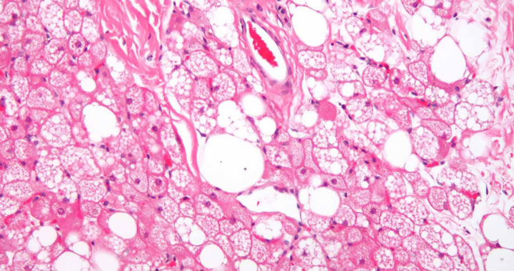
Brown adipose tissue and cells or brown fat is one of the three types of fat abundant in hibernating animals and newborn humans. Compared to white adipose tissue or white fat, which contains a single lipid droplet, brown fat contains numerous smaller lipid droplets and a higher number of iron-containing mitochondria. Note that beige fat shares characteristics with both brown and white fat. Scientists initially thought that brown adipose tissue had negligible function in adult humans. However, according to several studies, it plays an important role in metabolism, especially in the consumption of energy.
Brown Fat as a Solution to Obesity and Raising Metabolism: A Review of Studies
Understanding the Role of Brown Adipose Tissue
Brown fat is responsible for non-shivering thermogenesis. This process involves generating heat and raising body temperature by burning calories instead of shivering.
Researchers S. Rajakumari et al. explained that brown fat burns excess energy and white fat stores them. They identified B cell factor-2 or Ebf2 as a protein responsible for fat cell development, differentiation, and function. Their experiment showed the functional difference between white fat and brown fat by overexpressing Ebf2 in precursor white fat cells.
The induced brown adipose tissue consumed higher amounts of oxygen, had a greater number of mitochondria, and had an increased expression of genes involved in heat production.
It is worth mentioning that the capacity of brown fat tissue to generate body heat is about 300 times greater than other tissues in the body. This tissue is abundant in children but its amount decreases over time through adolescence and further into adulthood. Lindsay Robinson et al. explained that most adults only have 50 to 100 grams of brown fat.
Nevertheless, because brown adipose tissue or brown fat burns calories at rest, other researchers are investigating its potential in weight loss and in preventing obesity.
Combatting Obesity by Exploiting Brown Fat
Sugatani et al. discovered a link between a deficiency in the platelet-activating factor receptor or PAFR gene and the development of obesity due to impaired thermogenesis.
Specifically, after silencing the PAFR gene in mice, they observed that the subjects developed brown fat dysfunction. This meant that they were unable to effectively produce heat, had a reduced metabolic rate, and were more susceptible to gaining excess weight. This indicated that a dysfunctional PAFR gene results in brown fat dysfunction.
Researchers Laurie Goodyear et al. reiterated that the accumulation of white adipose tissue or white fat is associated with increased body mass and obesity.
However, because brown adipose tissue is associated with lower body mass index and high-energy consumption, they hypothesized that it has some role in maintaining a leaner and metabolically healthier phenotype. They also postulated that a brown fat tissue transplant could be a therapeutic approach to combatting obesity and other metabolic diseases.
A brown fat transplant in selected mice was performed. These mice were also fed with either a normal diet or a high-fat diet to test the hypothesis.
Recipient mice had improved glucose tolerance, increased insulin sensitivity, lower body weight, decreased fat mass, and a complete reversal of insulin resistance induced by a high-fat diet after 8 to 12 weeks. The transplanted brown fat also secreted several important hormones. These include the pro-inflammatory and anti-inflammatory interleukin-6.
Brown fat transplants could be a possible solution to manage weight and combat obesity in people struggling to lose weight despite following common intervention programs.
It is still important to underscore the fact that the separate studies of Rajakumari et al. and Junko Sugatani et al. also suggest the use of protein targeting through drug therapy Remember that Ebf2 was identified as a protein responsible for the differentiation of fat cells. This also includes the adequate functioning and maintenance of brown adipose cells.
The problem with this critical protein is that it cannot be readily targeted using drugs or with other conventional pharmaceutical approaches.
Researchers need to determine a workaround to effectively and efficiently express Ebf2 and turn white fat cells into brown fat cells. These could possibly include gene therapy, molecular tools for boosting its expression, and potential upstream regulators. Another option is to re-engineer white fat cells to behave like beige fat cells or brown fat cells.
The study of Rajakumari et al. noted that it is possible to pharmacologically block or stimulate the interaction of this protein with another partner protein.
Cold Climates and Brown Adipose Tissue
Other studies further suggest using natural mechanisms to stimulate the energy-consuming activity of brown adipose tissue or make brown fat cells more active.
Carpentier et al. enrolled six healthy adult men in a study that involved controlled cold exposure conditions. All subjects demonstrated substantial non-esterified fatty acid and glucose uptake upon cold exposure. The findings also demonstrated cold-induced activation of oxidative metabolism in brown fat but not in neighboring skeletal muscles and subcutaneous fat tissue.
Overall energy expenditure increased under cold temperature. However, in warm temperature, similar energy expenditure and activations were not observed.
The findings of Carpentier et al. are also echoed in another study by Paul Lee et al. that involved enrolling five healthy adult men and subjecting them to temperature acclimation that lasted for four months. Results revealed that long-term exposure to cold environments can stimulate brown fat growth and activity by about 30 percent to 40 percent.
It is also worth noting that prolonged exposure to warm climates or warm temperatures decreased the amount of brown adipose tissue below the baseline level.
Tore Bengtsson et al. explained how cold environments activate brown adipose tissue. The process involves the sympathetic nervous system activating adrenoceptors on the surface of brown fat cells when the body is exposed to cold temperatures. This activation stimulates the glucose uptake from the bloodstream. Brown fat cells use glucose as fuel to generate heat.
An accompanying commentary on the study of Carpentier et al. mentioned that increasing the amount of brown adipose tissue in a person is unlikely to make them leaner.
Impact of Stress on Brown Fat Activity
Stress also appears to induce brown fact activity. Researchers Labros S. Sidossis et al. studied burn trauma patients to investigate and understand this connection.
Collected samples of white adipose tissues from 72 patients who sustained severe burns revealed a gradual shift in the molecular and functional characteristics of white fat cells to a more brown fat phenotype over time. This suggested the progressive browning of white fat cells in response to a burn trauma and prolonged exposure to physical and biological stress.
Researchers Sidossis et al. further explained how stress can result in the browning of white fat cells. Note that brown fat cells express a protein called UCP1.
This protein prompts the mitochondria to burn calories without producing chemical energy. Burn trauma triggers stress and the release of adrenaline. The entire process activates UCP1. Sidossis et al. concluded that stress as a pathway for the browning of white fat cells is possible and their study can lead to the development of drugs that can mimic the effects of burn trauma.
Robinson et al. also showed that even mild stress from activities like a math test can activate existing brown adipose tissue and result in body heat generation.
FURTHER READINGS AND REFERENCES
- Rajakumari, S., Wu, J., Ishibashi, J., Lim, H.-W., Giang, A.-H., Won, K.-J., Reed, R. R., and Seale, P. 2013. “EBF2 Determines and Maintains Brown Adipocyte Identity.” Cell Metabolism. 17(4): 562-574. DOI: 1016/j.cmet.2013.01.015
- Robinson, L. J., Law, J. M., Symonds, M. E., and Budge, H. 2016. “Brown Adipose Tissue Activation as Measured by Infrared Thermography by Mild Anticipatory Psychological Stress in Lean Healthy Females.” Experimental Physiology. 101(4): 549-557. DOI: 1113/ep085642
- Stanford, K. I., Middelbeek, R. J. W., Townsend, K. L., An, D., Nygaard, E. B., Hitchcox, K. M., Markan, K. R., Nakano, K., Hirshman, M. F., Tseng, Y.-H., and Goodyear, L. J. 2012. “Brown Adipose Tissue Regulates Glucose Homeostasis and Insulin Sensitivity.” Journal of Clinical Investigation. 123(1): 215-223. DOI: 1172/jci62308
- Sugatani, J., Sadamitsu, S., Yamaguchi, M., Yamazaki, Y., Higa, R., Hattori, Y., Uchida, T., Ikari, A., Sugiyama, W., Watanabe, T., Ishii, S., Miwa, M., and Shimizu, T. 2013. “Antiobese Function of Platelet‐Activating Factor: Increased Adiposity in Platelet‐Activating Factor Receptor‐Deficient Mice with Age.” The FASEB Journal. 28(1): 440-452. DOI: 1096/fj.13-233262